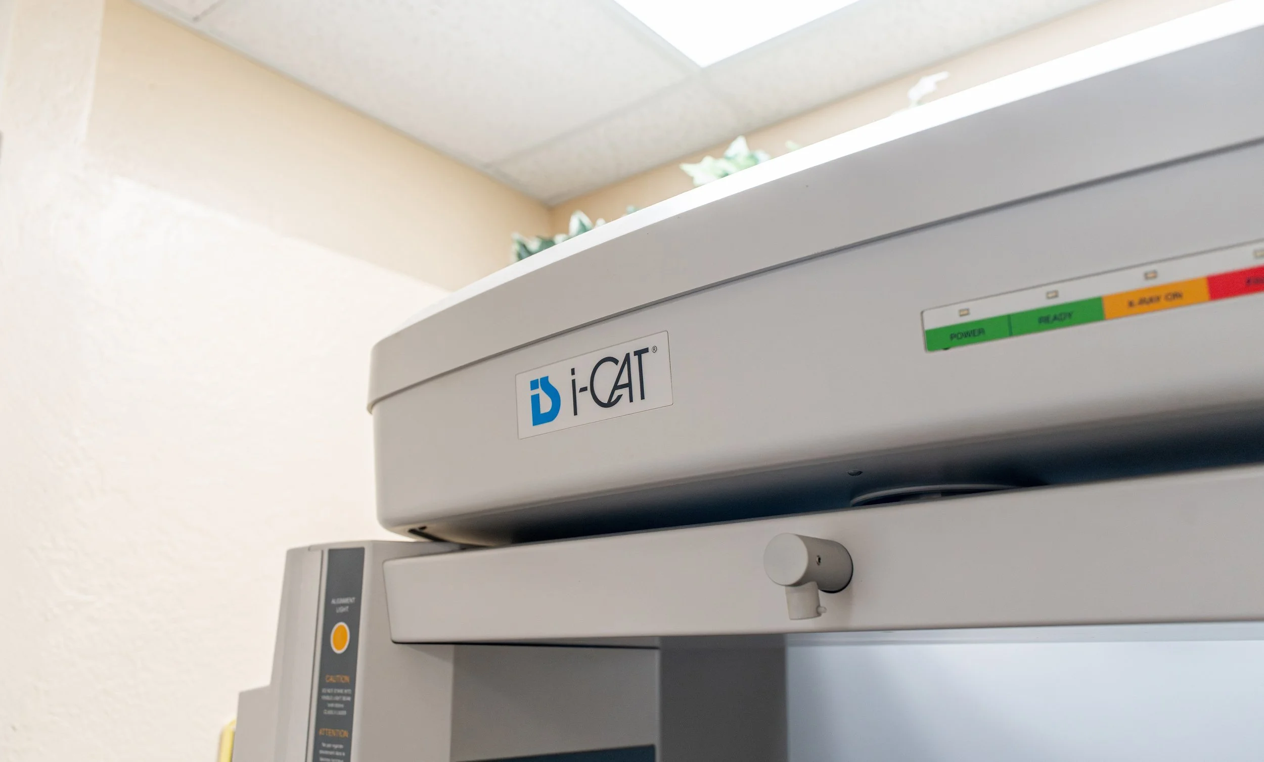
What is Cone Beam CT (CBCT)?
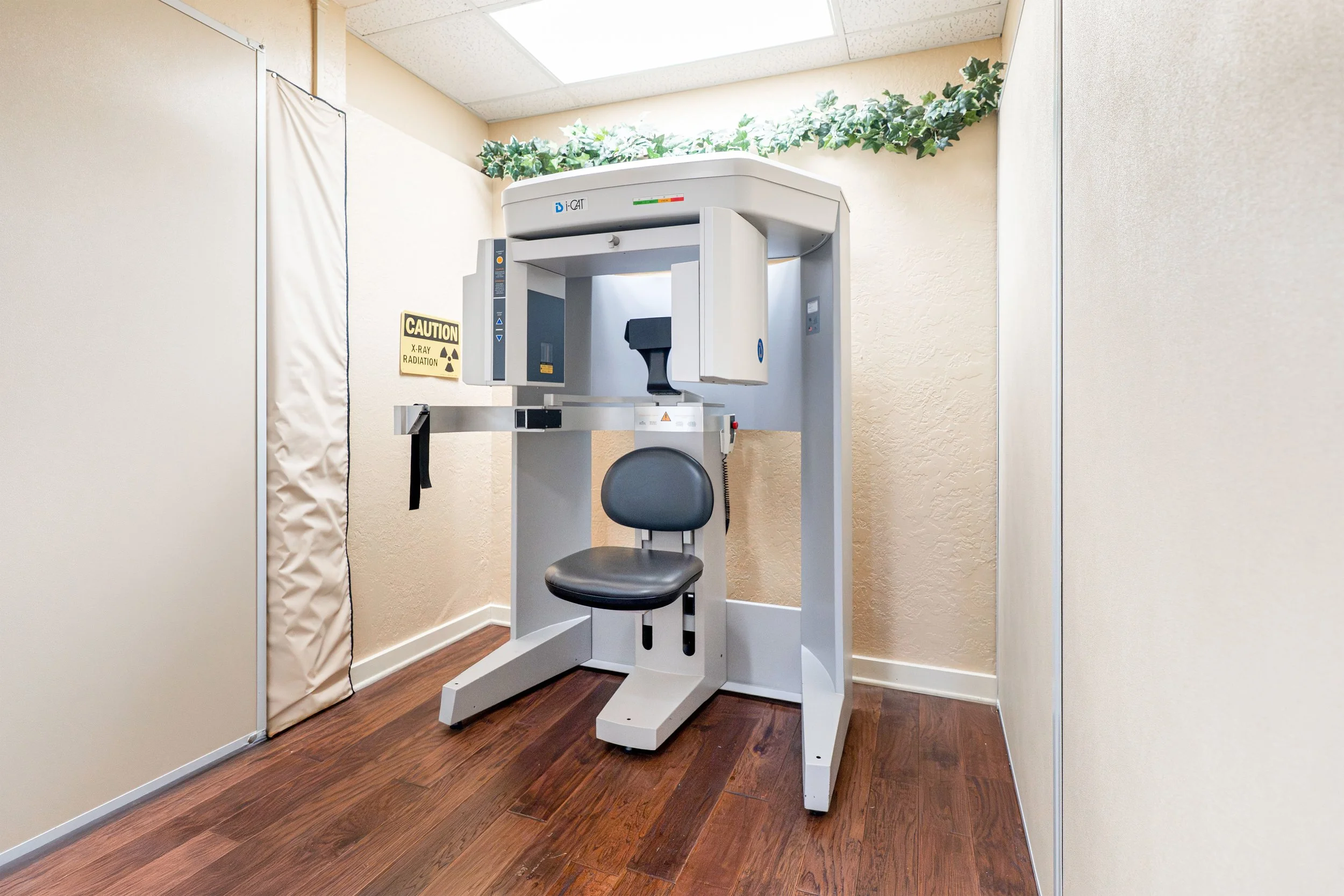
At the Head and Neck Center of Hawai‘i, we utilize Cone Beam Computed Tomography (CBCT), an advanced imaging technology that produces highly precise three-dimensional views of the head and neck. Widely trusted in dentistry for its accuracy, CBCT enables us to evaluate the atlas vertebra and surrounding structures in exceptional detail. By integrating this technology into Atlas Orthogonal care, we ensure the most accurate analysis and provide individualized treatment tailored to each patient.
Benefits of CBCT in Chiropractic
-
CBCT provides detailed three-dimensional images of the bony structures of the upper cervical spine, which is what the Atlas Orthogonal analysis requires. CBCT is not a substitute for MRI or conventional CT. When soft tissue evaluation such as disc or spinal cord assessment is clinically indicated, Dr. Perrino will refer you for the appropriate imaging.
-
CBCT images provide detailed information about the size, location, and shape of spinal abnormalities, allowing chiropractors to develop more effective treatment plans. For example, CBCT can help chiropractors determine the best location for spinal adjustments or identify areas that require additional therapy.
-
Traditional upper cervical analysis requires a series of separate x-ray views to capture the necessary detail. A single CBCT scan of the cervical spine provides three-dimensional detail at a radiation dose comparable to or lower than that multi-view series, and substantially lower than a conventional medical CT. We scan only the cervical spine rather than a larger field. Dr. Perrino orders imaging only when it is necessary for your analysis and will discuss that decision with you.
-
By providing more accurate diagnoses and treatment plans, CBCT can help improve patient outcomes, reducing pain and improving overall function. CBCT can help patients better understand their conditions and treatment plans, leading to increased satisfaction with their chiropractic care.
Cone Beam CT and Atlas Orthogonal Chiropractic
Head and Neck Center of Hawai’i utilizes specialized imaging called Cone Beam CT. Cone beam computed tomography (CBCT) is a specialized type of computed tomography (CT) that uses a cone-shaped X-ray beam to produce high-quality 3D images of structures inside the body, such as bone and soft tissues. In Atlas Orthogonal Chiropractic, CBCT imaging is used to create a detailed image of the upper cervical spine and surrounding structures. This allows Dr. Perrino to accurately assess the alignment of the atlas (C1 vertebra) and axis (C2 vertebra), which are the first two vertebrae in the neck. The Atlas Orthogonal technique involves the use of a specialized adjusting instrument to gently correct any misalignment in the atlas vertebra. By using CBCT imaging, Dr. Perrino can ensure that the adjustment is precise and targeted, as well as monitor progress over time. Overall, CBCT imaging is a valuable tool in Atlas Orthogonal Chiropractic as it provides detailed, accurate information that helps guide treatment and ensure optimal outcomes for patients.